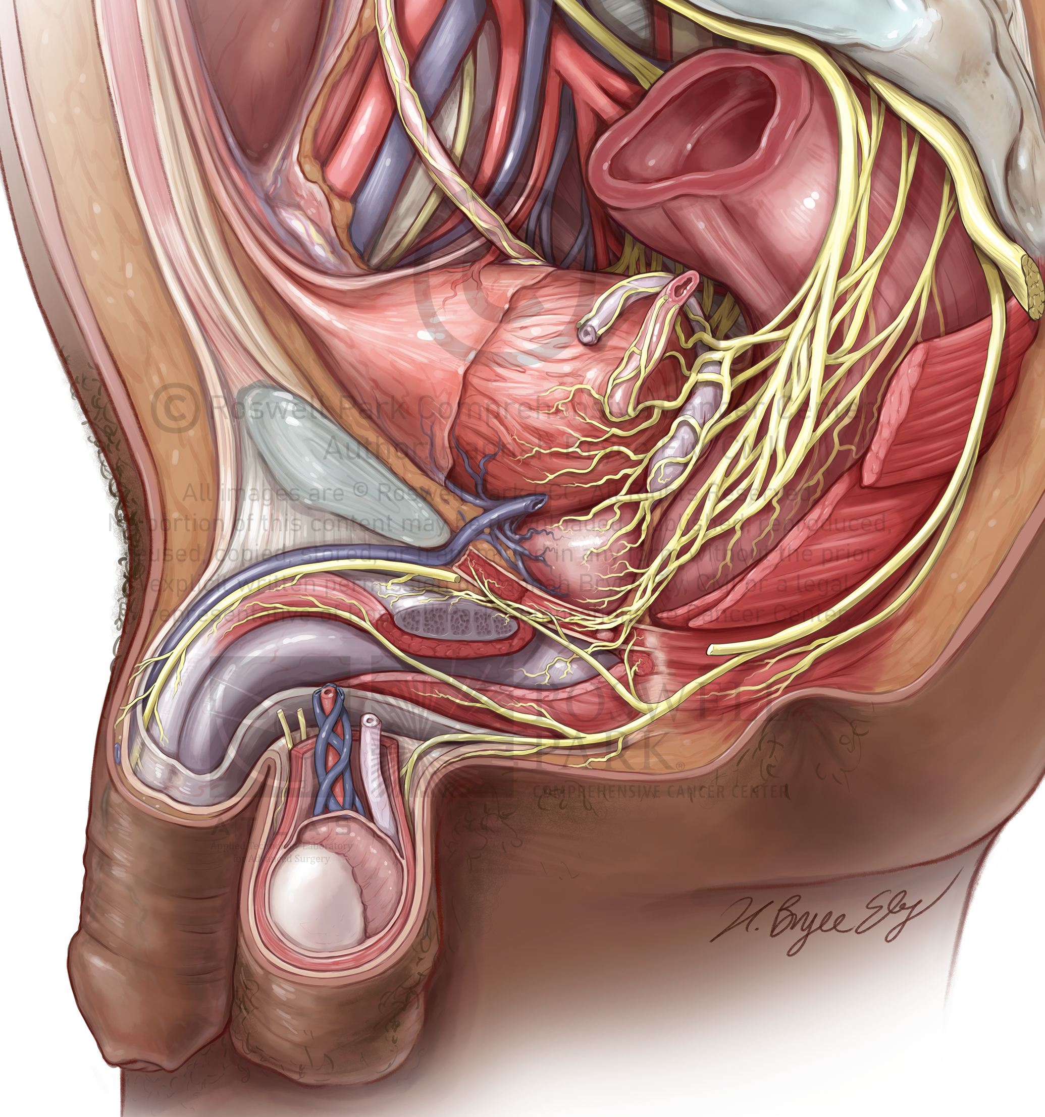
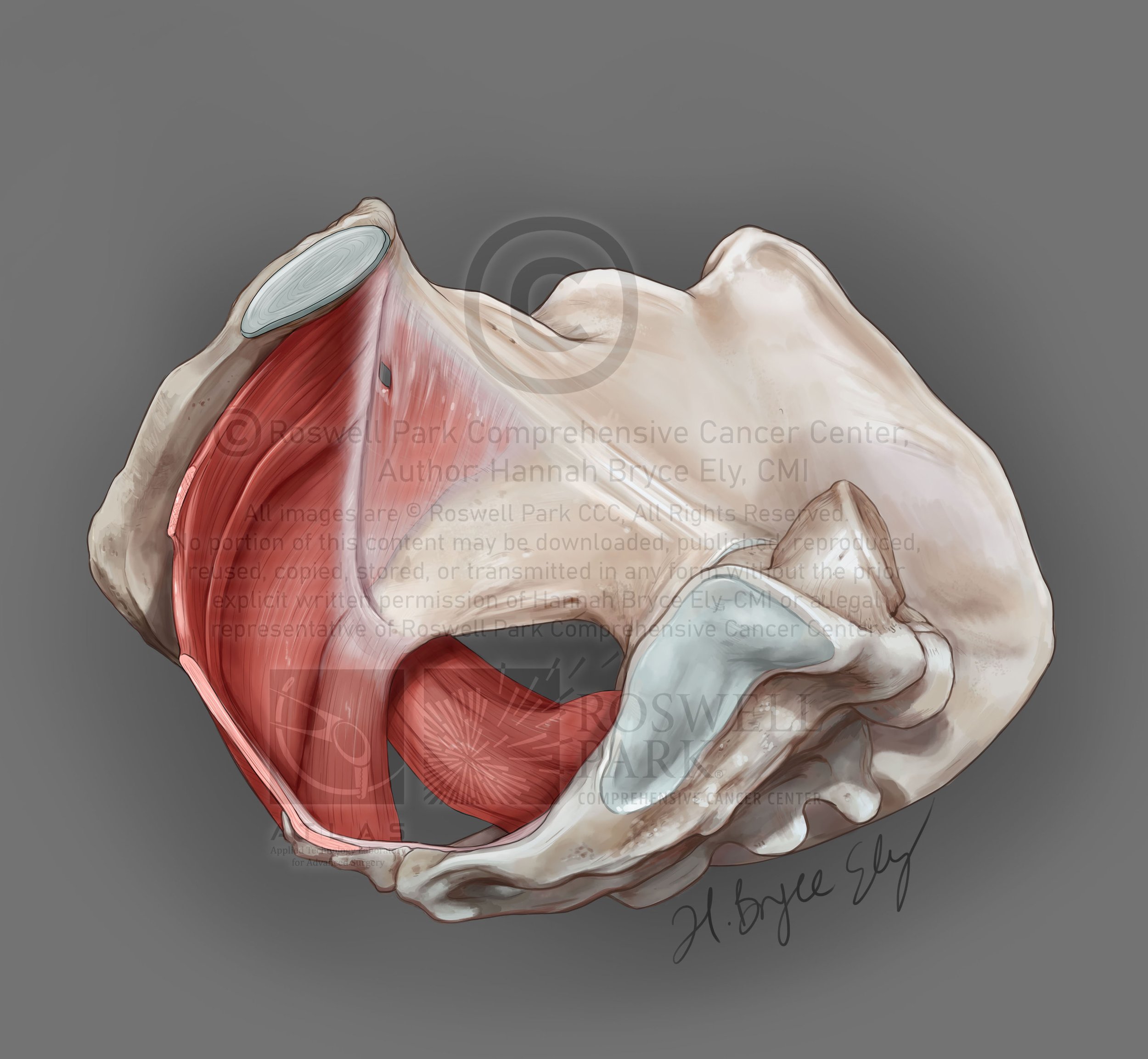
Pelvic nerve anatomy: inferior hypogastric plexus
The autonomic nerve plexes of the pelvis are difficult to define and visualize. These illustrations were created to clarify this difficult anatomy for training urologic surgeons. Injury to these nerves during pelvic surgery can cause incontinence and impaired sexual function in men and women, so understanding of these pathways is crucial for positive patient outcomes.

© Roswell Park Comprehensive Cancer Center, ATLAS


© Roswell Park Comprehensive Cancer Center, ATLAS

© Roswell Park Comprehensive Cancer Center, ATLAS

© Roswell Park Comprehensive Cancer Center, ATLAS

© Roswell Park Comprehensive Cancer Center, ATLAS

© Roswell Park Comprehensive Cancer Center, ATLAS
© Roswell Park Comprehensive Cancer Center, ATLAS
Nerve-sparing (NS) cystectomy is a treatment for bladder cancer. In this surgery, the bladder and other organs are removed and the autonomic nerves of the pelvic visceral plexus that supply erectile tissue and the external urethral sphincter are spared to preserve sexual function and continence. Historically, this procedure has been recommended for male patients. Now, more surgeons are attempting to bring NS techniques to female patients. The purpose of these illustrations is to provide detailed anatomy of the plexuses showing pathways of autonomic innervation from the SHP to the cavernous nerves and structures being innervated. The layout compares male and female anatomy, showing analogous structures and critical differences surgeons must understand as they perform NS more widely. Inclusion of the pudendal nerve and its branches shows overlapping somatic innervation. Because these nerves cannot be seen in the surgical field, surgeons must be confident in this anatomy. These illustrations—informed by several recent papers, cadaveric dissections, 3D modeled CT data, and review by expert surgeons—were created for a urologic atlas to educate surgeons on techniques, procedures, and surgical anatomy. Although male pelvic plexus anatomy is better studied and understood, the exact course of these nerves in male and female patients is yet to be confirmed. Giving surgeons this visual comparison is crucial for successful outcomes for all patients as NS technique is expanded.
These illustrations were shown as a layout in the Association of Medical Illustrators 2022 Salon, and received an Award of Merit.
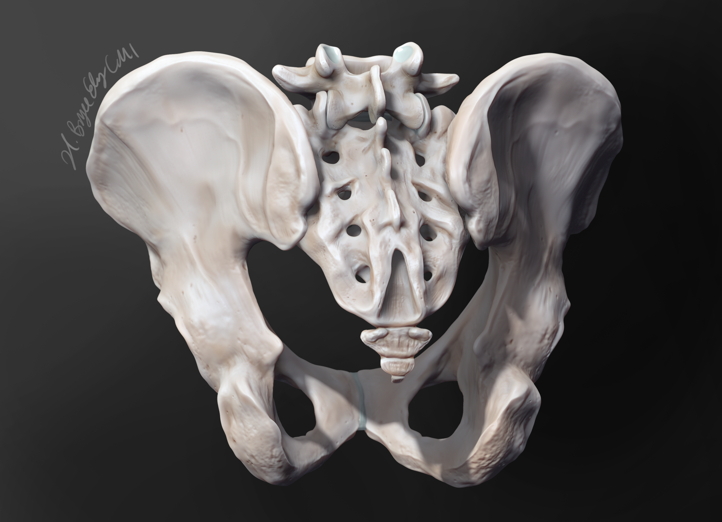
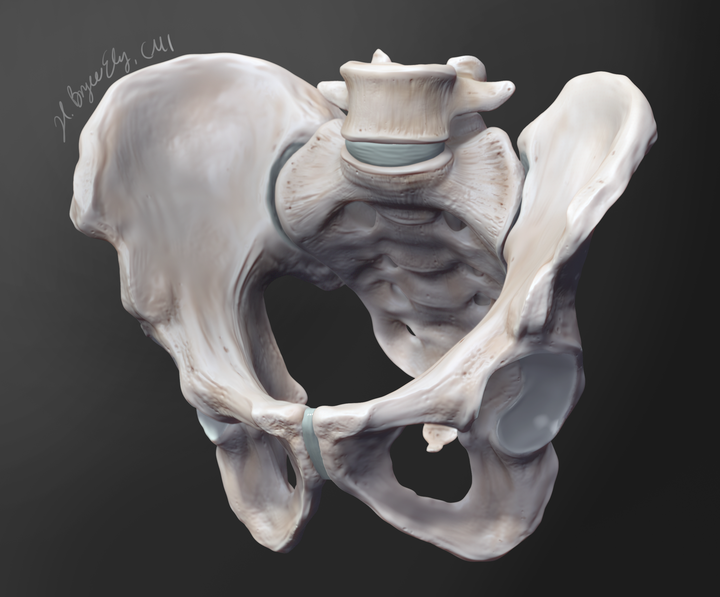
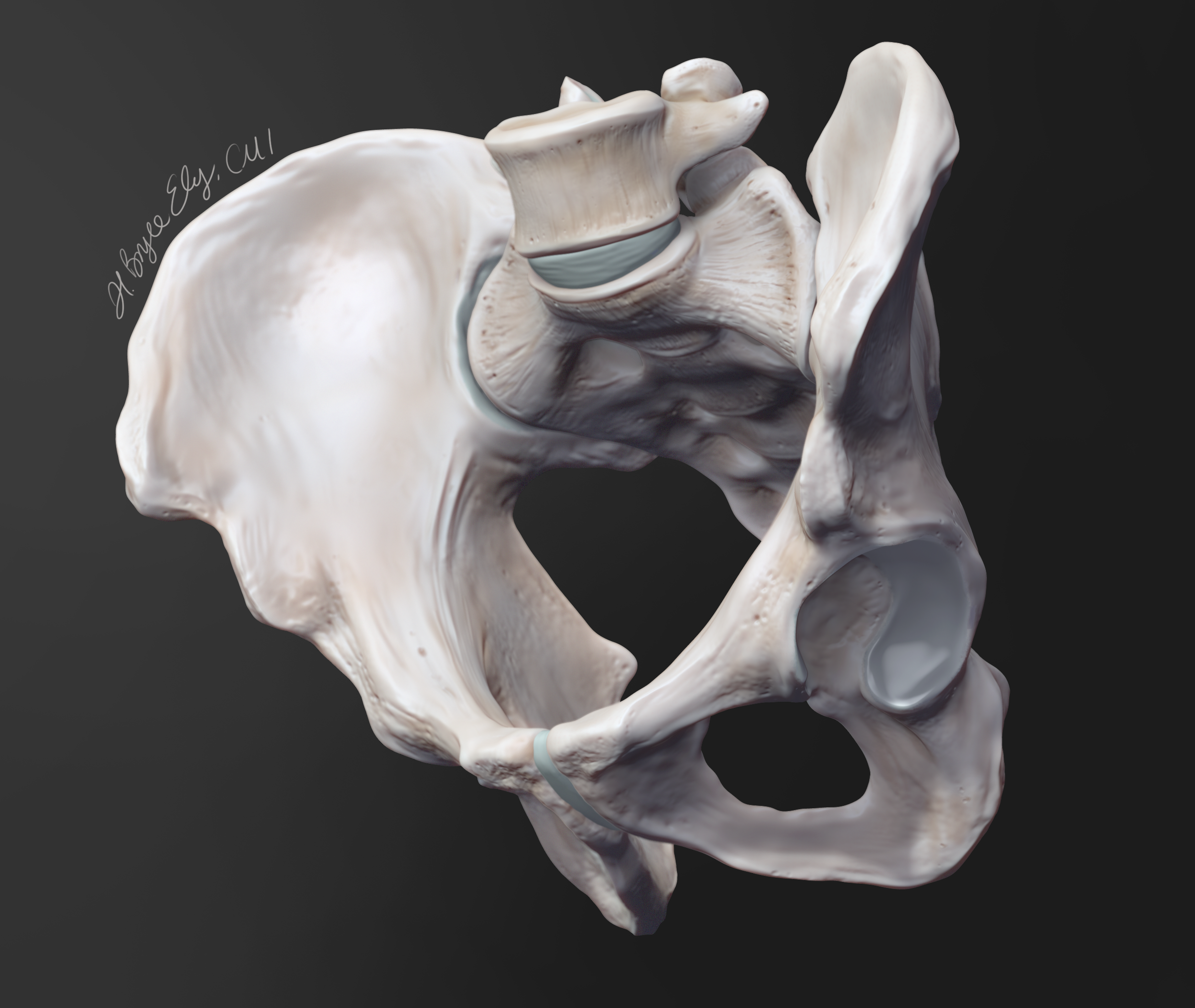
3D model created in ZBrush, in progress adding organs, nerves, and vessels. Female pelvis in progress as well.


This illustration was created for a chapter in Campbell-Walsh Urology. This shows the anatomy of the prostatic and vesical plexuses involved in nerve-sparing surgery. This image was used to alert surgeons and clinicians to the areas of common nerve injury during cystectomy, so that these injuries can be avoided during surgery.


Let’s Work Together
I welcome new projects and opportunities in the world of healthcare and science communication.